This is a fixed price procedure costing £3000. We offer a free initial consultation with our orthopaedic vet Kate Downie, to assess whether this procedure is the right fit for your dog.
Cruciate Ligament Disease in Dogs
There are two cruciate ligaments in the knee (or ‘stifle’ as we call it in dogs). Cruciate disease (usually the cranial cruciate ligament) is the most common cause of hindlimb lameness in the dog.
People often acutely traumatise their cruciate ligament while playing sports whereas in dogs the process is more chronic causing degeneration of the ligament over time.
Diagnosis
Usually, diagnosis is made upon clinical examination of your pet alongside radiographs (X-Rays) to confirm the diagnosis and also to assess for arthritic change.
Treatment
Some small dogs (less than 15kg) may do well with a period of rest and anti-inflammatories alone, although surgery is generally considered to offer a quicker and more reliable recovery.
Larger dogs are less likely to do well without surgery and so surgery is always advised. Numerous stabilisation techniques have been described.
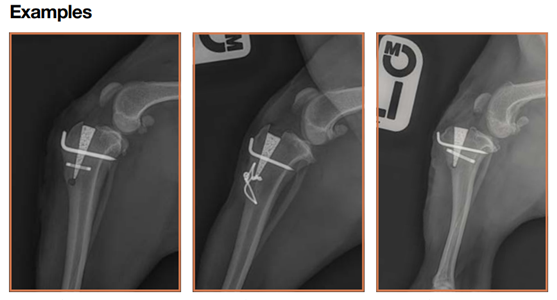
At Parkhill Vets we can treat your pet’s cranial cruciate ligament disease surgically using a geometry changing technique known as the Modified Maquet Procedure (MMP).
The Modified Maquet Procedure (MMP)
The Maquet Procedure (MP) was developed by orthopaedic surgeon, Dr P Maquet, in the 1960s to treat patellofemoral pain and knee osteoarthritis (OA) in people. The tibial tuberosity advancement (TTA) procedure is an evolution of the MP that was developed by Montavon, Tepic et al in 2002. The Modified Maquet Procedure (MMP) is an Orthomed modification of the TTA procedure used to treat cranial cruciate ligament (CrCL) deficiency-related lameness.
The MMP uses a wedge-shaped implant of OrthoFoam™ which both defines the degree of advancement of the tibial tuberosity and holds the bone in its new place while the bony ingrowth that provides permanent, biomechanically robust fixation develops. The use of a carefully engineered saw guide ensures a controlled, precisely directed and positioned osteotomy of the correct length, resulting in an appropriately “thick” tibial tuberosity while at the same time protecting adjacent structures from iatrogenic injury.
The open porous structure and sympathetic mechanical characteristics of the OrthoFoam™ wedge provides robust early and sustained bony ingrowth fixation obviating the need for bone grafting, postoperative support bandages or a lengthy period of restricted activity. The potentially disruptive forces that act to displace the distal end of the tibial tuberosity cranially following advancement are controlled using either a tension band wire or a titanium staple.
We are always happy to discuss options with owners and offer referral where appropriate.